Anatomy Chest Muscles Diagram / Thorax Anatomy Wall Cavity Organs Neurovasculature Kenhub - Human anatomy diagram shoulder anatomy shoulder muscles shoulder muscles and chest.. Want to learn more about it? Almost every movement in the body is the outcome of muscle contraction. Muscles, connected to bones or internal organs and blood vessels, are in charge for movement. The chest anatomy includes the pectoralis major, pectoralis minor and the serratus anterior. Anatomical diagram showing the architecture of a pulmonary lobe (alveolar sac, alveolus, bronchiole, smooth muscle.)
Now that you understand about what muscles make up your chest, their function, location and the rep range needed to stimulate them, let's give you some workouts to help you build. The chest wall is comprised of skin, fat, muscles, and the thoracic skeleton. Their main function is contractibility. Attached to the bones of the skeletal system are about 700 named muscles that make up roughly half of a person's body weight. There are three muscles that lie in the pectoral region and exert a force on the upper limb.
Adduct Extend And Rotate For Better Pecs Breaking Muscle from cdn3.omidoo.com Note that the middle lobe. Muscles, connected to bones or internal organs and blood vessels, are in charge for movement. In this article, we shall learn about the anatomy of the muscles of the anterior chest. The pectoralis major muscles (also known as the pecs) are located on the front of the rib cage, and form the major muscles of the chest. A massive chest anchors the upper body and enhances the appearance of your shoulders, arms, and abs. Related posts of chest muscles diagram. Anatomy of the chest and the lungs: Anatomical diagram showing the architecture of a pulmonary lobe (alveolar sac, alveolus, bronchiole, smooth muscle.)
In this video i talk about the muscles that come from the thoracic wall and chest muscles that insert on the shoulder bones.✅.
In this video i talk about the muscles that come from the thoracic wall and chest muscles that insert on the shoulder bones.✅. The pectoralis minor muscle (not shown in the diagram) is located underneath the pectoralis major muscle, attaching to the coracoid. Related posts of chest muscles diagram. They are the pectoralis major, pectoralis minor, and the serratus anterior. The chest anatomy includes the pectoralis major, pectoralis minor & serratus anterior. The pectorals, or chest muscles, are so large and prominent that they can't be hidden. Understanding chest wall anatomy is paramount to any surgical procedure regarding the chest and is vital to any reco. Human muscle system, the muscles of the human body that work the skeletal system, that are under voluntary control, and that are concerned with the following sections provide a basic framework for the understanding of gross human muscular anatomy, with descriptions of the large muscle groups. The interactive muscle anatomy diagram shown below outlines the major superficial (i.e. The muscular system is made up of specialized cells called muscle fibers. There are around 650 skeletal muscles within the typical human body. Almost every movement in the body is the outcome of muscle contraction. Located immediately below the skin) muscles of the body.
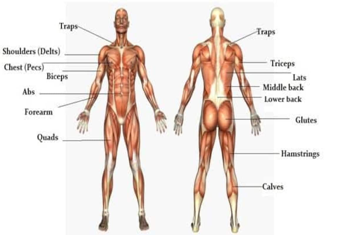
The shoulder muscles bridge the transitions from the human muscle system, the muscles of the human body that work the skeletal system, that are under voluntary control, and that are concerned with movement, posture, and balance. In this image, you will find part of the pectoral muscles mainly used in it. The pectoralis minor muscle (not shown in the diagram) is located underneath the pectoralis major muscle, attaching to the coracoid. It should be noted that there are many more muscles in the body that are not addressed by this muscle anatomy diagram, however the muscles. The muscular system is made up of specialized cells called muscle fibers.
Muscles Of The Chest And Abdomen Kirtley S Anatomy Website from kirtleycaldwellanatomy.weebly.com Located immediately below the skin) muscles of the body. In this article, we shall learn about the anatomy of the muscles of the anterior chest. The chest anatomy includes the pectoralis major, pectoralis minor and the serratus anterior. All about the chest muscles. This is a table of skeletal muscles of the human anatomy. Learn the anatomy of the shoulder muscles now at kenhub. They are a gland, so there is a sources (like lingerie websites) only feature chests that display their products to their best advantage. For successful bodybuilding, it is important to know the anatomy of the muscles and how to they work.
Want to learn more about it?
Learn about each of these muscles, their locations, functional anatomy and exercises for them. Their main function is contractibility. Free online quiz back and chest muscle diagram. Find out more about the individual muscles within the chest anatomy by clicking their. Diagrams showing the general organisation of the thorax with the pleural cavity and lobule: In this post, you will learn the chest muscles anatomy which is easy since there are not so many muscles. Attached to the bones of the skeletal system are about 700 named muscles that make up roughly half of a person's body weight. Adducts & flexes the arm (humerus). Human anatomy diagram shoulder anatomy shoulder muscles shoulder muscles and chest. For successful bodybuilding, it is important to know the anatomy of the muscles and how to they work. The chest anatomy includes the pectoralis major, pectoralis minor and the serratus anterior. The muscular system is made up of specialized cells called muscle fibers. Learn about each muscle, their locations & functional anatomy.
Male digestive system diagram 2021 | male and female digestive system anatomy anatomynote.com found chest muscle anatomy from plenty of anatomical pictures on the internet. Tough connective tissue at the bottom of the calf muscle merges with the achilles tendon. Related posts of chest muscles diagram. Free online quiz back and chest muscle diagram. Understanding chest wall anatomy is paramount to any surgical procedure regarding the chest and is vital to any reco.
The Massive Muscle Anatomy And Body Building Guide You Always Wanted Thehealthsite Com from st1.thehealthsite.com Their main function is contractibility. In the diagrams below, i'll be showing muscle groups in color, with a black line to show the forms first a few words about anatomy: Name and locate major muscles of the human body on a torso or diagram. Attached to the bones of the skeletal system are about 700 named muscles that make up roughly half of a person's body weight. It provides protection to vital organs (eg, heart and major vessels, lungs, liver) and provides stability for. Related posts of chest muscles diagram. The shoulder muscles bridge the transitions from the human muscle system, the muscles of the human body that work the skeletal system, that are under voluntary control, and that are concerned with movement, posture, and balance. The chest wall is comprised of skin, fat, muscles, and the thoracic skeleton.
Their main function is contractibility.
1300 x 1390 jpeg 297 кб. Learn about each of these muscles, their locations, functional anatomy and exercises for them. Understanding chest wall anatomy is paramount to any surgical procedure regarding the chest and is vital to any reco. Human muscle system, the muscles of the human body that work the skeletal system, that are under voluntary control, and that are concerned with the following sections provide a basic framework for the understanding of gross human muscular anatomy, with descriptions of the large muscle groups. Anatomy • free medical books. The chest wall is comprised of skin, fat, muscles, and the thoracic skeleton. All about the chest muscles. The chest anatomy includes the pectoralis major, pectoralis minor and the serratus anterior. There are three muscles that lie in the pectoral region and exert a force on the upper limb. Diagrams showing the general organisation of the thorax with the pleural cavity and lobule: Trapezius icon vector from anatomy collection. Related posts of chest muscles diagram. Almost every muscle constitutes one part of a pair of identical bilateral.
Diagrams showing the general organisation of the thorax with the pleural cavity and lobule: chest muscles diagram. The chest anatomy includes the pectoralis major, pectoralis minor and the serratus anterior.